December 2025
Rise in Dinoflagellate and larval forms
What a change this month. November was dominated by typical winter diatoms (Odontella and Bacillaria) with few, if any, dinoflagellates. The trend continued at the start of this month. The sample taken 23rd December was an absolute cracker! Phytoplankton components have switched positions with now a low diatom count but the highest I have ever recorded for dinoflagellates. Usually the most I see in the x100 microscope field of view is one or two but now, two days before Christmas, 5 or 6 together. Plus, 10 different species were common, even a couple of Noctiluca. I have never seen so many dinos in one sample. I use a 53 micron mesh net but really a finer mesh (25 micron) is better for a good sample of dinos. This makes my haul of dinoflagellates especially astonishing. See the photos to see the most abundant species.
It seems like spring has come early because I had been seeing dinos going into resting stages to pass the winter back in November. None were present on 23rd just a sudden surge in Tripos, Dinophysis and even Protopteridinium. The later tend to be a spring/summer genus. P. pentagonum, a kidney-shaped individual is not one I often see, particularly in the middle of winter, and it was quite abundant. So what is going on? We had been having strong winds early in the month with the Haven very churned up. This would have brought nutrients as well as resting stages off the bottom up into the light. Dino resting cysts need several months before “germinating” so these must have been cysts from late summer, I am suggesting. This is the first time I have ever had such a density and variety of species together. Amazing!
Diatoms are a golden yellow and related to kelps and other brown seaweeds. By contrast it can be rare to find a green single-celled member of the marine phytoplankton. One of these is Halosphaera, a green alga, that stands out. They were abundant on the 23rd.
Gyrosigma

Two species of Tripos together, T. furca and T. tripos

Tripos fusus

Three Tripos individuals amongst cyphonautes larvae of sea mat
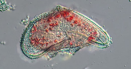
Protopteridinium pentagonum
This sign of spring has also occurred in the zooplankton. The small periwinkle Melaraphe has been releasing eggs rather early, since late autumn. These were common in the first week of the month but only one specimen occurred on the 23rd. Instead, huge numbers of mollusc larvae, both bivalves and snails. Perhaps the most dominant larval type was of Bryozoa or sea mats, the cyphonautes larva. These can live for several weeks in the plankton and occur, typically, over the winter period. I always see them about now but never in the numbers I saw on the 23rd. They must have made up more than half of the samples by volume, with 6 or more present in the microscope field of view. Normally it would be one.
Strangely, parasites were abundant on 23rd, more so than usual. At least three different cercaria larvae occurred. These are of parasitic flatworms (think liver fluke) and pass through molluscs to birds and fish. So common were they that several would be present together under the microscope. I species was so large they were almost visible to the naked eye. A group of non-parasitic flatworms, the marine Polyclads, have larvae one of which is called a Muller larva that live for a short time in the plankton. Several appeared in my 23rd sample. A favourite of mine they are very active and have eight pod-like extensions covered in cilia. They are a delight to watch. Back in August the larva of the tiny isopod (crustacean like a woodlouse) of the family Bopyrid were very common. The larva, called a cryptoniscus, parasitises copepods is surprisingly common at the moment.

The bioluminescent dino Noctiluca

The green alga Halosphaera is unusually common

Odontella sinesis the most common diatom at the moment

Cyphonautes larvae

Out-stretched cercaria with sucker in the centre

Bopyrid cryptoniscus larva

A pair of contracted cercaria of the family Lepoceadiid
There is currently a sizeable surge in larval polychaete worms of all stages from the trochophore with several bands of cilia through to larger forms with the characteristic bristles, called nectochaete larvae. There was no Tomopteris (the “elegant worm”) like last month, a worm that lives permanently in the plankton, but there was another strange worm that I am still trying to identify. At the moment I think it is Pelagobia but I really not sure and need to investigate further. It is only a millimetre or so long and seems to have a well developed gut.

A large trochophore larva of a marine worm

The head of a possible Pelagobia worm

A Muller larva

One of many Sabellaria worm trochophores present this month

A young Idotea isopod
To top off the Christmas excitement in the latest sample there were young Idotea (isopods) and several gorgeous oppossum shrimps, Mysids present. These were juveniles about 8 – 10 mm long. Happy New Year!
November 2025
Gelatinous Plankton!
Gelatinous plankton was high this month. To begin with there was the appearance of very young jellyfish, the Mauve Stinger Pelagia noctiluca. Early in the month a sample off Skomer secured several individuals between 1 and 3 cm across. By 23rd November several late stage ephyra larvae were present in the Dale collection - the first time I have seen it here. Pelagia has been a rare occurrence, especially in the Haven, because it is an Atlantic species that moves and reproduces around the ocean. Unlike our other jellyfish species it is not tied to the shore by a polyp stage attached to the shore as it is entirely adapted for a pelagic life. Unusually for a jellyfish, the sting cells (nematocysts) are found all over the body including the surface of the bell or umbrella, visible as small warts. The flat, blade-like oral tentacles as well as the long thin tentacles both collect zooplankton that is then wiped across the mouth. On the various photos here note the small brown structures called rhopalia that are sense organs detecting gravity and light. The photo of the juvenile in the blog news is a different individual to the Photo of the Month specimen and is older so that brown coloration is beginning to occur; mauve appears later with maturity.
The siphonophore Muggiaea has all but disappeared from the Haven but is still common off-shore.

Juvenile Pelagia noctiluca; note the warts of sting cells on the top
Gyrosigma

Pelagia: a single oral arm and tentacle. The arrow indicates a rhopalium, sense organ

The beautiful polychate worm Tomopteris helgolandica

Parasagitta elegans, an arrow worm (Chaetognatha) appearing this month in the Haven
Other zooplankton considered to be examples of gelatinous plankton are the transparent permanent species like the polychaete worm Tomopteris helgolandica and arrow worms. The former is one of my absolute favourites and occurs occasionally across the winter into the spring. It was exciting to find a mature adult in the sample, just over 20 mm long. Like other “jellies” they can be extremely difficult to see as they are almost completely transparent. It was only when magnified and I saw a swirl in the water, catching a glimpse of the tips of some parapods (side extensions), I realised it was there. They are particularly tricky to photograph. Autumn is also a time for arrow worms to appear in the Haven, common off-shore but not always in the Dale sample. They are formidable hunters of copepods using their bristle jaws and sharp teeth to grasp the prey. The individuals that turned up this month were all juveniles around a centimetre in length.

Late ephyra lava of P. noctiluca
Once the small jellyfish were in a dish a dozen or so small crustaceans began whirling around in the water. These were a species of Hyperia, an amphipod crustacean that is parasitic on jellyfish. At only a few millimetres long I was surprised as the species shown elsewhere (see parasitic crustaceans) on this website is H. galba and significantly larger at around 12 mm. There were several females with young and is probably H. medusarum.
Another crustacean that was fairly common this month was a Podocopid ostracod, which first appeared last month (see the October photo below). Marine ostracods are found in freshwater and out in the oceans but not often in seashore plankton. These are individuals that live in the surface of sediment at the bottom and have been either disturbed or swum up to join the plankton (called tychoplankton).
From the end of October large numbers of eggs have been shed by adults of the small periwinkle Melaraphe neritoides which live on the jetty where I collect samples. They have so much yolk around the egg that the embryo doesn’t need to feed in the plankton, just be dispersed.

Corethron

Juvenile Hyperia medusarum, a parasitic amphipod

Egg of the small periwinkle Melaraphe neritoides
The phytoplankton is going through a slow decline but there is a good variety and density of diatoms present. Odontella species dominate along with the sliding diatom Bacillaria paxillifer, all species I consider to be winter plankton. I was pleased to see these being joined by the lovely Corethron hystrix, again a species I see often during the winter. Pleurosigma and Gyrosigma were both common this month and like the ostracods probably washed off the sediment. It has been rough this month and all that turbulence has created a wealth of foraminiferans to be present. They are a type of amoeba that secretes a tiny shell in which it lives. The small holes on the shell surface allows the pseudopodia (cell extensions) to pass through to grab food. With all the rain and snow there has been flooding and high volumes of water in the estuaries. This has brought down a freshwater desmid Staurastrum that was in very dense numbers at the end of November. The fact that they have chloroplasts suggest that they have not been in the sea for long. Dinoflagellates have been declining with a good deal of resting spores, especially of Polykrikos (see last month) but Dinophysis caudata that I only recoreded for the first time last month is still present.

Podosira, a tiny but common diatom in November

The freshwater desmid Staurastrum

Pleurosigma


Two examples of many different foraminiferans common this month

Is this oddity a sporangium of a fern?
Weird oddities appear every month and often I fail to identify them in the debris. More than one of these strange objects (pictured left) appeared. This was the most complete example and the only thing I can think of is that this is the sporangium of a fern. Found under the fronds these release spores. The tough spine (top and right side) is the annulus that dries out to propel the spores in to the wind. Particularly odd when I have never seen one in plankton before but in one sample several turned up.
October 2025
Dominance of Phytoplankton
The zooplankton has been diminishing with the first lack of echinoderm larva since the spring. By contrast the phytoplankton has exploded in numbers and diversity. Rhizosolenia robusta, a very large diatom has suddenly become very common and the tiny Cerataulina pelagica is still abundant although now in longer chains. One that comes and goes through the year but is producing large numbers of resting spores is Biddulphia alternans. The genus name has several synonyms where taxonomists cannot agree and I have written the one I can remember(!). R. robusta is also a recent name change, Neocalyptrella was the previous one. For the last six months Thalassionema has ben seen occasionally but has become common around the Haven this month. Quite small it has to be magnified over x400 to see the detail. Gyrosigma requires less magnification and has become abundant especially around Neyland, in the eastern Haven. Here the salinity is lower than Dale and the water depth is less above the sediment. It is a benthic species living in the surface of sandy mud. Down the length of this diatom is a raphe system of canals that use exuded mucus to move through the sediment. This is typical of the pennate type diatoms that dominate the benthos and others species are common in the plankton here like Navicula. True planktonic species are typically the centric forms and do not need to move to find light.
In July dinoflagellates were in abundance and have continued through into the autumn; then we had Storm Amy early in October. Afterwards, Tripos species, if they occurred were damaged with horns missing. Otherwise, they disappeared. Strangely, two new species suddenly turned up a week after the storm. I have never seen Dinophysis caudata here before but it has become common in Dale. (compare with others from July). The other was a species of Polykrikos. I find resting stages of these on occasions but rarely good specimens.

Rhizosolenia (Neocalyptrella) robusta

Gyrosigma

Biddulphia alternans, resting spores

Thalassionema sp.

Damaged Tripos missing horn

Dinophysis caudata

A pseudocolonial dinoflagellate Polykrikos
Polykrikos is a type of dinoflagellate that is often found in small colonies called pseudocolonies, in the above case of 8 zoids. They feed on diatoms and other dinoflagellates.

Dinoflagellate Noctiluca with tentacle extended
Colonial diatom, Thalssiosira subtilis. has been common this October
I have reported for a few months now that the harpacticoid copepod Euterpina acutifrons has been abundant in the plankton. It is one of the few harpacticoid types that lives pelagically and not in the sediment. There is a diatom Sceptronema orientale that is highly specific to growing attached to the copepod and became a common sight on the copepod.
Diatoms are a golden brown colour, affirmation that they are brown algae related to kelp. When a green alga occurs, uncommon in marine plankton, it shows up well. Storm Amy created a strong downpour of rain that washed a number of freshwater algae down the estuaries into the Haven. One I have not seen for a while, Pediastrum, is a particular, beautiful favourite as it produces lovely colonial forms. I think this is P. duplex, common in lakes up in the Preselli Hills. Like the freshwater desmid, Staurastrum, that is very common in the Haven, the cell walls are especially tough and resist decay and can exist for years after death. The fact that the Pediastrum is so green suggests they have been freshly brought down by the river.
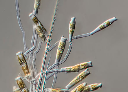
Sceptronema orientale, a diatom specific to growing on the copepod Euterpina

A small colony of Pediastrum, a freshwater alga
Tintinnids are a form of ciliate, single-celled protists. Abundant in marine plankton they have had a succession of population explosions since the spring (see the various species in the monthly blogs below). In the last two months there has been a new dominant genus Leprotintinnus. All tintinnids secrete a lorica (a protective case) in which it lives. Some are like wine glasses while others like this species are tubes open at both ends. Eutintinnus has a clear tube and appeared in June but Leprotintinnus studs the lorica with minerals as can be seen in the photo here. The ciliate lives at one end of the open tube with the crown of specialised cilia propelling it through the water as it collects food to engulf. The samples this month had high numbers of this species.

The ciliate Leprotintinnus at the end of it's lorica


The siphonophore Muggiaea with the copepod prey
The siphonophore Muggiaea atlantica is a summer and autumn regular visitor, multiplying offshore where it is abundant and moving into the Haven with the tides. They feed on copepods and often I find one trapped by the colonial polyps of Muggiaea, located in a small pocket at the base of the gelatinous bell. In the second photo here you can see a close-up of the calanoid copepod with the feeding polyps at the top that have stung and immobilised it. Most exciting is that the poor copepod already had problems as on its back there is an ectoparasite in the form of an isopod crustacean that has specialised to attach and feed on them. A third photo shows a close view of this larval Bopyrid isopod that are very difficult to identify and separate to species. Back in August the numbers of Bopyrids swimming in the plankton was high but I have never had such a good view of one actually on a calanoid copepod.

Ectoparasitic Bopyrid isopod
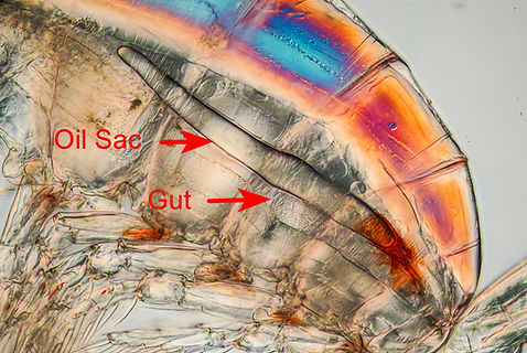
Copepod Calanus showing oil sac

Caligus
Copepods have been in quite high numbers all summer coming into autumn and as part of their preparation for winter they store a waxy lipid in their oil sac which lies above the gut. In the photo this stands out well along with the red pigment at the posterior end of this sac. This is probably to reduce the presence of sunlight passing out of the oil sac end which could attract fish predators from below as the sac acts like a lens focusing the light from the surface. During winter the calanoids go into deep water where they diapause, a form of hibernation.
Early autumn is when I always find a few Caligus copepods, an ectoparasite typically on on fish. At 5 mm or so they are distinct in any sample and look to me like baby flatfish swimming around the dish. More here on Caligus. Another parasite, the monstrillid copepods, have been present since the spring but now have finally disappeared.
Corycaeus is from a family of predatory copepods that have huge eyes, like a pair of headlights. I didn't see them last year but in 2023 they appeared in the autumn. This october they arrived offshore in large numbers anmd some entered the Haven occurring in a few samples. I find the eye fascinating and the photo shows a stacked image of the complex structure x400 magnification. Behind the lens is a crystalline cone that focuses the light on to a light sensitive rhabdome (like a retina). The specimen shown also has a diatom growing attached to the lens surface. It looks suspiciously like Sceptronema, see above, that is supposed to be specific to Euterpina.

The eye of Corycaeus

Podocopid Ostracod
And finally, a bit of a surprise was another crustacean, a podocopid ostracod. they look a bit like the cyprus larva of a barnacle but smaller. I only find them very occasionally and this is not typical plankton but has come from the sediment at the bottom. Rather nice though.
A sample taken today at Dale was a bit of a surprise as a large influx of arrow worms has occurred from offshore. But more of that next month ...
September 2025
Diatom Blooms and Metamorphosis
Autumnal changes begin in August. Biodiversity was increasing, especially in the gelatinous plankton with Sarsia the dominant hydromedusa. Different diatoms of the phytoplankton were on the increase, and chains of Chaetoceros curvisetus were the most abundant species present this month. The large Coscinodiscus diatom species have been fairly absent all summer but now their numbers are almost as dominant as Chaetoceros species. A notable diatom, one of my favourites that I call the "sweet wrapper" due to the twists in the chain, suddenly appeared in reasonable numbers: Helicotheca tamesis.

Long chains of Chaetoceros curvisetus

Helicotheca tamesis

Coscinodiscus sp.
The numbers of dinoflagellates in the phytoplankton was surprisingly low. Tripos and Dinophysis species were replacing the spring Protopteridinium forms in July when there had been an explosion of dinos (see July below). However, Tripos has been in fairly low numbers. Then at the start of September Noctiluca scintillans became common in the Haven to be easily the most abundant dino species present. This is typical for the last few years: common off-shore throughout the summer and then entering Milford Haven in the autumn. An intriguing species that is a predator hunting with a tentacle.

Noctiluca x100. Note the expanded tentacle
Noctiluca scintillans

Noctiluca scintillans
The harpacticoid copepod Euterpina acutifrons was an abundant copepod last month and continues to be with many carrying eggs. These eggsacs could be found floating free in the plankton and several were hatching into tiny nauplii larvae.
Eggsac of Euterpina with hatching nauplii

Eggsac of Euterpina with hatching nauplii
The nauplius is the characteristic larval form of copepods and Cirripedia (the barnacles). The latter have some specialised species such as the endoparasite Sacculina carcini which has a very complex life cycle using the host the Common Shore Crab Carcinus maenas. The nauplius is quite distinct and was prolific in the plankton at the end of August running into September.
.jpg)
Euterpina acutifrons with an eggsac

A nauplius of the endoparasite Sacculina carcini
Ophiopluteus metamorphosing into a juvenile brittlestar

Ophiopluteus metamorphosing into a juvenile brittlestar

The larvae of brittlestars, called an ophiopluteus, have been common both in the Haven and off-shore in St Brides Bay through the summer. In early September larger individuals could be seen metamorphosing into tiny juvenile brittlestars. The latter continued to appear through the month.
Sea squirt tadpole larva. NB. the yolk cells attached to the body
Juvenile Brittlestar

Juvenile Brittlestar
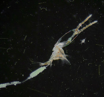
Monstrillid, the semi-parasitic copepod
Several species of Monstrillid copepods have been common in the plankton since May but this is the first month there has ben fewer present. It has been amazing seeing these semi-parasitic species, not least because I have never seen them occurring in the Haven before. Their nauplii quickly become endoparasitic on gastropods and polychaetes living on the shore while the adults disperse but do not feed reliant on food built up during the larval phase (see photo of food stored in the legs and lack of organs in the anterior body). It has given me an opportunity to take plenty of photos.

Monstrillid head
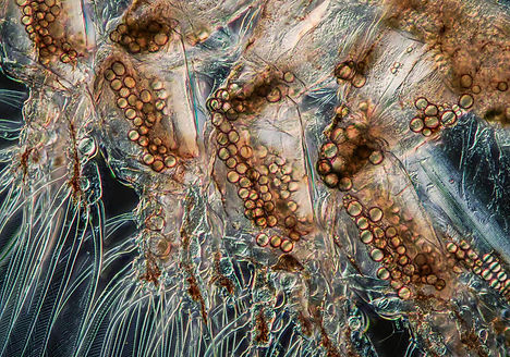
Monstrillid Legs showing lipid storage
August 2025
Late Summer Blooms

A tintinnid "murmuration"

Tintinnids magnified x200
Hydromedusae
There are invariably microscopic "jellyfish" in samples, just the odd one or two, invariably of the hydroid Obelia, which is found as a colony of polyps on the rocky shore. The medusa is the dispersal and sexual phase of the colony. Called a hydromedusa, as they are specific to hydroids, this month there was a large increase but of a different genus, Sarsia. Dozens of examples were found in the Dale samples. A sample from off-shore near Skomer was absolutely heaving with Sarsia, probably S. gemmifera. The hydromedusa of this species produces buds from the "stem" inside (the manubrium) which develop into new medusae: medusoid buds. Obelia medusae are shed from the polyps.
The ciliate group Tintinnina have been abundant since spring but this month in the western Haven they have exploded in numbers. The sample looks to the naked eye like it is full of sand. Allowed to settle they swarm around a dish like microscopic murmurations constantly changing shape. Quite mesmerising. The width of the photo is 1 cm. Under the microscope many of them were conjugating, where they "swap" genes.
As well as an increase in these single-celled animals an increasing diversity of diatoms are beginning to bloom. The tiny Ceratulina (blooming in May) is back. Striatella unipunctata is a diatom species found on rocky shores but has suddenly become abundant in the plankton. We have had some windy periods and water turbulence has washed the diatoms off the shore. The chain diatom Chaetoceros curvisetus is blooming and is the most dominant species.

Ceratulina (left) and Striatella

Chaetoceros curvisetus

Sarsia medusoid buds x200
Polychaete worm larvae have been abundant this summer and the most dominant species has been Lagis (Pectinaria) koreni. The adult is common, living in a beautiful tube in the sand in the lowest areas of the shore. The young larva contracts back inside a "hood" but when expanded moves rapidly through the plankton.

The hydromedusa of Sarsia with a number of medusoid buds

Young polychaete larvae of Lagis koreni, contracted and expanded


Adult Lagis koreni. Head view showing golden chaetae

Euterpina acutifrons
The harpacticoid copepod Euterpina acutifrons is the dominant copepod species. Most harpacticoids live in sediment but this is one of the few that is truly planktonic.

Bopyrid isopod crustacean

Bopyrid isopod crustacean
Another common crustacean this month is a tiny isopod, a Bopyrid, that parasitises the outside of copepods. I have never seen so many in the plankton.

Different views of an early actinotroch larva of Phoronis.

I think the expression is that "they are like buses, you wait for ages and several come at once". Maybe one or two Phoronid larvae appear a year but since June every sample has several. These actinotroch larvae are all early forms, around 100 microns in size, with between two and eight developing tentacles (more like ciliated pods). As they grow and develop in the plankton this number increases, up to 40 or more. After 3 weeks they are 1 - 2 mm in length. See photo of the month.
Sea squirts or Ascidiacea that live on the shore release eggs in the summer that hatch into a tadpole larva. Both were common this month. The larva does not feed in the plankton but lives off yolk cells that persist after hatching.

Sea squirt tadpole larva. NB. the yolk cells attached to the body

Sea squirt egg with developing larva
July 2025
Dinoflagellate Blooms
This year has been full of surprises: a succession of different blooms and new species. Last month ended in low diversity but high in the quantity of copepods, polychaetes and tintinnids. There are still plenty of marine worm larvae but the copepod population has virtually disappeared. The phytoplankton was very low, difficult to find a diatom, but that has begun to change. Usually a collapse in diatoms results in the tunicate Oikopleura increasing and that is so. At the start of July the plankton was absolutely dominated by their pale gelatinous bodies, which you can see in the sample photo. It was probably the highest density of these I have ever seen. They are perfect fish food and it was noticeable that as I sampled so I could see shoals of large fish moving in with the tide to feed.

The green Halosphaera and tintinnid lorica.

Low magnification view of a sample. Huge numbers of the white tunicate Oikopleura, tintiniid "shells" (ciliates) and cockle larvae.

Cockle veliger larvae with tintinnid "shell" or lorica.

Protoperidinium, common in previous months producing a resting cyst inside, 24th July

Tripos tripos, now appearing. This one is dividing (mitosis) to create two new daughter cells, 24th July.

Dinophysis acuta, with the two flagella just visible (red arrows)

Dinophysis tripos
The tintinnids, a form of ciliate (single-celled protist) that secretes a shell called a lorica around it and then studs it with minerals, are still abundant. The lovely Favella shown in the photos last month have all but disappeared so the group are dominated by small oval ones of probably 3 or 4 species. Stenosemella is probably the most common genus present. These along with cockle veliger larvae fill in the gaps between the Oikopleura.
The dinoflagellates have been represented by Protoperidinium, the "spring genus", so far this year (see previous months) but now all change. I have never seen it so clearly marked as one population disappears another rises and takes over. As can be seen in the two photos the outgoing Protoperidinium is dying and making a resting cyst inside. Meanwhile the sudden rise of Tripos species (4 so far in the last sample) has been dramatic and many were dividing to asexually reproduce. They split across the centre and each daughter needs to replicate the other complex half. The genus Tripos was called Ceratium but now that name is only used for freshwater species.
The dinoflagellate bloom is quite spectacular because not only are there several Tripos species blooming but several other genera. I have occasionally seen Dinophysis acuta and D. tripos before in small numbers but never at the same time. The former is very common at the moment with the latter in smaller numbers. And a new species for me: Phalacroma rotundatum, one of the smallest here at just 35 microns. Quite tricky to photograph as it whirled around and one of the flagella can be seen. Dinos have two flagella and these are just visible in the D. acuta photo (red arrows). The two Dinophysis species are about 50 microns long.

Phalacroma rotundatum

The protist Acanthometra

A copepod resting egg

A Muller Larva of a Polyclad flatworm
These were some of the highlights but plenty more interesting organisms are turning up. It follows on from the comment at the end of June in that the late summer/early autumn blooms are already happening. Acanthometra is loosely considered a radiolaria, a fascinating cell (a bit like an amoeba) with strontium spikes passing through it. Often common in September and October, it is blooming now. The disappearance of copepods has been followed with the large number of resting eggs or cysts that see them through environmentally stressful periods. The active Muller larvae of the flatworm group, Polycladida are always great to see. never in large numbers because they exist for just 24-48 hours before becoming juveniles. I will finish on this new copepod species: Monstrilla helgolandica. It is an endoparasite on polychaetes and gastropods of the shore. The nauplii larvae are the parasites and on metamormophsis become non-feeding adults that disperse in the plankton. The anterior of the body is a void lacking organs. The photo below is just one of three examples found. Note that back in May I found a different species of the Monstrilloid group. See May below and also photo of the month back in May.
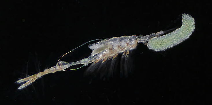
Monstrilla helgolandica
June 2025
Polychaete and Tintinnid Blooms
The last few months have been about successive phytoplankton blooms but at the start of June it was marine worms or polychaetes that completely dominated samples. The delightful little Polydora larvae were prolific and I have never seen such concentrations of them at Dale, only in the trapped water of the Gann Lagoon where larvae are common in the sediment. All larval stages were present. The tiny individuals that have just developed chaetae/bristles only have small pouches for palps. As they mature extra segments are added at the posterior end creating longer larvae with larger and larger palps.

Low magnification view of a sample showing polychaete worm larvae mainly Polydora

Magelona showing the very long bristles.

A different view of the head of Magelona with contracting tentacles

Tomopteris, a worm that lives permanently in the plankton.
In addition to Polydora was another Spionid larva that is quite rare in Dale shore samples and several examples were present: Magelona mirabilis. The adult is around 10cm long and lives in clean sand in the sublittoral. The extended and flattened head gives the common name of shovel-head worm. Although I have occasionally looked I have never found them at the bottom of Dale's sandy shore but it is wonderful to see these larvae. Just a few millimetres in length they have two very long extended palps and many long chaetae. All of these along with the palps will help to keep the animal drifting in the plankton, reducing the chance of sinking. NB. the best shot is photo of the month.
It was also super to find several of the transparent Tomopteris worms (my favourite) and I have to photograph them despite possessing numerous photos. These were juveniles, just over a centimetre in length. Adults are two or three times the size. The yellow spots produce bioluminescence.

The polychaete Poecilochaetus.
This is a young worm, called a nectosoma, approximately 3mm long
A really unusual worm that I have never encountered before is the polychaete Poecilochaetus. It too is a form of Spionid. The larva is largely transparent except for the area around the parapodia and where bristles attach. Little is known about the adult except that it lives in sediment. By all accounts the larva lives in the plankton for a long time. The young planktonic worm is called a nectosoma and less than half a centimetre long. June is usually a quiet month so finding so many worms has been a treat.
Polychaetes were not the only zooplankton that bloomed. The ciliate group, the tintinnids, proliferated in huge numbers. When this happens it is usually one of the small species with the lorica, within which it lives, studded with minerals. Instead there was a massive rise in the transparent loricate forms like Favella and Eutintinnus. I have never seen a bloom like it. This coincided with a drastic reduction and death of phytoplankton that had been blooming from March until mid-May. Possibly, the rise in decay and minute particulate matter enabled the filter feeding ciliates to increase.
With the large numbers of Favella present I was able to find for the first time some that were parasitised. Euduboscquella is a dinoflagellate genus that lives inside the tintinnid cell where it produces spores that disperse to parasitise other Favella individuals. The parasite lifecycle is complex and varied with them producing different kinds of spore or sporocyte. Some large "macrospores" build up inside the lorica and a number of individuals like this were found. A few had chains of "microspores". These sporocytes disperse and infect fresh hosts where they are ingested and live for 4 -6 days before more sporocytes form. These cells have a pair of flagella and in the smaller ones they could be seen jiggling about together inside the lorica. In most cases the host seemed to be still alive.
When I first saw the sporocytes I initially thought it was to do with the tintinnid reproduction. Upon checking recent tintinnid literature this would appear to be how they were originally classified 100 years ago. In more recent years the complex cycle of these dinoflagellates is beginning to unravel.

Favella, a tintinnid with a transparent lorica (case or test)

Eutintinnus, with a tube lorica, open and both ends

Favella with the lorica full of large sporocytes of the internal parasite dinoflagellate Euduboscquella

Euduboscquella inside Favella
End of June: A sample taken on 27th June confirmed the polychaete and tintinnid numbers were still very high with Tintinopsis now the dominant species. Overall, the general biodiversity of the plankton was declining (but only slightly) but the density of species was very high. Previous June samples have shown a real drop in both volume and biodiversity; a different trend this year. The appendicularian/tunicate larvae had reached a peak and the number of developing cockle eggs was high. Occasionally, I see nudibranch larvae but they were in great abundance as was the water flea Podon after disappearing from samples last month. All very strange. Most years I see the steady rise of groups like flatworms in August/September but it was great to see many different juvenile stages including several Muller larvae (Polyclad flatworm larva) which spend only days in the plankton. When it came to diatoms the weirdest feature is that there was a complete disappearance in all but three species, all of the genus Rhizosolenia, that was creating a massive bloom at the end of the month. The exception is Pleurosigma making its first appearance of the year. It tends to be a summer species here.
This has been an amazing month mainly because of its unpredictability and I cannot wait for July samples.

Tintinnopsis

Sporocytes of Euduboscquella dinoflagellate parasite
May 2025
A Massive Change in just a Few Weeks
At the end of April there was a diatom bloom but in the space of a few weeks the bloom shifted dramatically from medium sized diatoms to tiny ones and flagellates including Protoperidinium (dinoflagellates) and colonies of Phaeocystis globosa. It maybe worth having a quick look at April in the section below for comparison. The photo at the top of this blog shows a Comb Jelly or Sea Gooseberry (Ctenophore) Pleurobranchia pileus. This one was taken at Neyland but they are common at the moment throughout the Haven and is about a centimetre long. The water around the animal is thick with phytoplankton. A quick look at the seawater anywhere in the Haven and it looks scummy, slightly soupy and quite smelly.

Comb Jelly Pleurobranchia plieus amongst the huge phytoplankton bloom
The most dominant diatom in the phytoplankton bloom is a tiny diatom, Cerataulina pelagica. I have never seen a bloom of this before but is well known to do so, worldwide. It is well adapted to bloom quickly as it has a very thin, soft cell wall. Identification requires checking small, stumpy processes at the corners but as it rolls around they quickly disappear. The photo with red arrows show these at x400 magnification but only at over x1000 could I really see them. These weak processes and a small spine help to connect with others to produce short chains although I have not seen more than two joined together. Almost certainly the recent warm and very sunny weather has encouraged both this and Phaeocystis to bloom. Interesting, the diatom is only really blooming in the Haven while the Phaeocystis is more abundant on the open coast.

A single Cerataulina cell showing processes x1400

Two Cerataulina diatoms connected x1000

A nauplius trapped in a large gelatinous colony ofPhaeocystis globosa.

The dinoflagellate Protoperidinium currently blooming with the rest of the phytoplankton

Cerataulina pelagic a small diatom that has a soft and weak cell wall. Red arrow indicates the stumpy processes that connect cells
Due to the extreme density of the phytoplankton and "stickiness" of the mucus-producing Phaeocystis the bloom has limited and suppressed the zooplankton, particularly in the sheltered areas of the Haven on the eastern side, like Neyland. Even Dale has seen a lowering of the fauna. Larvae, like nauplii and those of polychaetes, are struggling although adults of larger species are doing alright. The sea gooseberry or comb jelly Pleurobranchia pileus has suddenly appeared and is strong enough to push through. Large copepods seem fine as well. The Photo of the Month is a species I have never seen here before, one of the parasitic copepods belonging to a family, the Monstrillids. In fact, in the same sample were 2 different species.

A clearer photo of Pleurobranchia, a comb jelly, common at the moment
Monstrillids are a group of endo-parasitic copepods but only in the larval phase where the nauplius lives inside the host, a gastropod mollusc or polychaete worm. The adult that emerges is non-feeding with no gut and lives for a very short time in the plankton, making it a rare find. The large eyes are not compound but more like mirrors and the flash used in the photo above has been reflected. The eggs are attached to spines instead of the usual arrangement of being in a sac.

The tunicate, Oikopleura

A late bipinnaira larva of a starfish

Cymbasoma rigidum female with eggs. Note the mirror eyes

A possible Monstrilla longicornis male
There had been a steady increase in Oikopleura during April but they can be inhibited by phytoplankton blooms. Although comparatively large (around a millimetre) they are not strong swimmers. It is no surprise, then, that their normal population growth seems to have stalled this month. They are an essential food source for fish in the summer when copepod populations can be low. We will see over the coming months if densities improve. However, a surprising turn is that for the first time I have seen both water flea genera, Evadne and Podon in Dale at the same time and in quite high numbers. Neither have been common in the Haven. Initially, they were parthenogenetic females but males have now appeared. This is to sexually produce offspring capable of surviving changing conditions. The Skomer marine reserve team provided a sample on 18th May where an Evadne "winter" egg occurred - my first photo of a resting egg. These can survive through difficult situations and can last for many years lying on the bottom. These are more typically found in freshwater water fleas and called ephippia. Information on marine forms are poor to non-existent but I am fairly sure this is what it is and it will be looking out for these in subsequent Haven samples.

Evadne from Skomer sample showing a potential resting egg inside

Evadne nordmanni

Podon
April 2025
Huge Diatom Bloom - Explosion of Polychaetes
At the end of March there was a huge increase in phytoplankton. This is almost entirely diatoms Although there were some Protoperidinium (dinoflagellates) present. With the good weather NASA acquired some brilliant, almost unique, images of the British Isles clear of cloud and displaying the phytoplankton bloom around the coast, especially around Pembrokeshire and Irish Sea. The very pale off-shore patches are coccoliths (haptophyte algae like those found in chalk). I never see them near the shore but a close relative is Phaeocystis globosa and that is common at the moment in shore plankton.

Plankton Bloom 7th April.
Photo credit: NASA Earth Observatory

The short chain diatom Lauderia annulata with external tubules

Long chains of the tiny diatom Skeletonema costatum x400
The diatom bloom consisted of just a few main species including the tiny diatom Skeletonema costatum. This has not been that abundant in recent years but Lauderia annulata is and often blooms in spring within the Haven. Thalassiosira gravida (it was rotula) also very common suddenly exploded in numbers this month. The genus Chaetoceros have "hairy" cells chained together (reduces sinking) and five species were common including C. socialis.
The polychaete trochophore larvae mentioned last month have developed into nectochaetes where the body is elongating as segments are added with more and longer bristles (chaetae). Most are Spionids or Polydora types with the prominent palps on the head. The larvae can be found throughout the year but are particularly abundant at this time of year.

Chaetoceros socialis chains

The dinoflagellate Protoperidinium with Thalassiosira gravida

Phytoplankton Sample dominated by Thalassiosira and Lauderia

One of many Polydora nectochaete larvae common this month.

Lovely to see the appearance of a number of pluteus larvae, in this case one of a sea urchin. A trochophore is caught up between two arms.

Synchaeta, marine rotifer female with egg

Barnacle Cypris larva
The abundant barnacle nauplii larvae last month have resulted in a mass of cypris larvae. Consisting of two valves it does not feed but contains large amounts of stored lipid (visible as round drops). The eye is the red spot. Protruding from between the valves (left) are the legs while the antennae at the front are sensory for detecting a good space to settle on the shore.
Additional crustacean larvae this month consist of zoea of both crabs and Galathea (Squat Lobster).
The rotifer population (Synchaeta) is developing in Dale. These are parthenogenic females and a large egg can be seen inside. Plenty of eggs in the water

Harpacticoid copepod Porcellidium, common this month

The rising numbers of the planktonic tunicate Oikopleura have stalled. This often happens when rapid diatom blooms occur. I think this is a female (gonad on top of head). I find them most abundant June - August after the spring diatom blooms.
March 2025
The Recovery continues now with Dinoflagellates
By mid-March there were dinoflagellates appearing, especially Protoperidinium species. Diversity of zooplankton larvae continues to expand. Nauplii of both barnacles and copepods had increased from last month. I have never seen so many crustacean zoea larvae in a haul and as well as crab zoea I had a very nice squat lobster Galathea zoea. In fact it was going to be my photo of the month until 15th March when I found a first for me: an antipathula larva. They are an exclusive larval form of the tube anemones. It is most likely Cerianthus lloydi that lives in a mucus tube in the mud. Looking like a toy rocket the four ciliated "pods" of the antipathula were slowly propelling it through the water when it stopped and gradually expelled what I believe to be old larval tissue through the slit mouth. The new slimline form then, over several hours, extended and increased the number of pods that will become tentacles in the juvenile anemone. The antipathula initially develops from a planula larva (typical of anemones) and I reported their sudden appearance last month. Planula numbers are currently higher than I have ever seen! See last months photo.

Squat lobster Galathea sp. zoea

Early antipathula larva of a tube anemone, mouth below

Later antipathula larva of a tube anemone, mouth above

Bryozoan coronate larva, 80 microns
Coronate larvae of certain species of Bryozoa last month but now prolific in numbers. Probably Bugulina sp. They only spend a matter of hours in the plankton before settling on the shore.

The dinoflagellate Protoperidinium blooming this month
The polychaetes have suddenly been releasing huge numbers of eggs and trochophore larvae. Probably the most prolific species is a sedentary spionid worm that lives in the middle of the seashore in mucus tubes within crevices and sediment, Malacoceros fuliginosa. The occasional egg is found but this month I was amazed at the number of them and plenty undergoing development into trochophores. The egg is distinctive in having a honeycomb envelope with specialised vesicles around the edge. Even the early trochophore larva has this honeycomb edge to it.

Egg case with developing embryo of Malacoceros, a polychaete worm
The phytoplankton is gradually increasing in diversity although abundance is still low. The sliding diatom Bacillaria paxillifer, which I always consider to be a winter diatom, is on decline while Odontella sinesis is about stable. Chaetoceros and Thalassiosira rotula are appearing in reasonable numbers. Corethron hystrix has always been a favourite species and is turning up in some numbers.
As expected, after several months of absence the dinoflagellates are back - resting stages from late autumn will be decysting. Protoperidinium is a spring/summer genus and is dominant. Tripos (formerly Ceratium) is abundant later in the year although a few T. furca and T. fusus have made an appearance.

The armoured dinoflagellate Tripos furca

The trochophore of Malacoceros. The arrows show where the ciliary band (prototroch) has formed.
February 2025
Rapid Recovery but still no Dinoflagellates
After a period of almost two months with the lowest diversity I can remember the first sample of February was amazing with more than double the number of species and a bloom of the large diatoms. By mid-February even smaller diatoms like Thalassionema and Chaetoceros were appearing. Most exciting is the presence of seashore larvae. Among the increasing numbers of barnacle and copepod nauplii larvae were those of sea anemones. One of my favourites to find these planulae are super to watch as the cilia are manic along the body but they just slowly glide around. Long sensory tufts at the front wave around. However, perhaps the most interesting to see was the sudden appearance of coronate larvae of a bryozoan species (sea mats or moss animals). These tiny hollow balls of cilia, 50 micron diameter, do not feed but spend just a matter of hours in the plankton before settling on the shore. Another bryozoan larva type called a cyphonautes was also abundant. This was probably the sea mat Membranipora. As they remain in the plankton, feeding and dispersing over a week or so they are commonly found unlike the coronates.

Planula larva of a sea anemone.
Note the tiny dots in the water, nanoplankton
Perhaps one of the most striking zooplankton species this month were the large number of Rhabditophoran flatworms that manically moved around the sample. They are so fast. The photo here shows some of the beautiful waves of cilia that moved across the body, just showing up along the edge. Approximately 450 microns when extended. They typically have 4 eyes but I am intrigued by what appears to be another two at the back! As carnivores they are feeding on the small zooplankton larvae. I am always amazed to see them swimming in the plankton as you might imagine that they need to be on a substrate like those living in freshwater or on the rocky shore.
Related to the Bryozoa is the phylum Phoronida. Very occasionally I find a young larva. On 13th February two were in the net. They must have just been released by the parents because in the first 24 hours they develop many lobes, mine barely had two. They grow into an amazing and large larva but by the time they get to this they have already been swept away by the tide. Interesting that this actinotrocha larva as it is called, along with coronates, are released at dawn. The low light on the water is the trigger. Too much light and the parents stop the release. In the case of the coronate larva it is to make sure the larva has chance to settle by the afternoon.

Bryozoan coronate larva, 80 microns
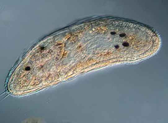
Rhabditophoran flatworm

Bryozoan cyphonautes larva
Other common larvae include the veligers of the Edible Periwinkle Littorina littorea. During January eggs of the small periwinkle Melaraphe neritoides were common but they have tailed off replaced by the larger periwinkle species. Melaraphe has so much yolk in the cell the larva spends only hours in the plankton while the L. littorea is around for a week or so. Greater chance of dispersal as it feeds in the plankton.

The complex actinotrocha Phoronis larva

Veliger larva of an Edible Periwinkle, common this month.

Larva of the polychaete Sabellaria with serrated bristles. Also common this month.

Several zoea larva of the Common Shore Crab
January 2025
Very Low Numbers So Far This Year
Although there has been a week of calm weather prior to the last plankton sample as well as resting stages of phytoplankton there was an amazing array of foraminiferans (a kind of amoeba living in a shell). They are benthic, living in the surface of the sediment but after a bit of disturbance can live in the plankton for some time until they settle again. I do not think I have ever seen so many as there were this time.
Happy New Year! Sampling for the last four weeks has been difficult due to poor weather conditions at suitable collection times. Generally, though, the plankton has been very low in diversity and density. A plankton collection in a sheltered area of the Haven near Milford at the start of the year was almost non-existent. Storm Darragh back in December decimated the plankton and recovery is going to be slow. The resting stages of dinoflagellates are still present and these will remain dormant until early spring. During the last 4 weeks I found just one living dinoflagellate. Diatom numbers are also still very low with the sliding diatom Bacillaria paxillifer being the dominant species although one could hardly say it is a bloom. This is unusual as I believe in the Haven there is always plenty of plankton to see at any time of the year with distinct winter species. A quick look at the 2024 blog at this time shows a reasonable biodiversity, then. On my scoring system this is the lowest value I have recorded in 4 years. Both common Odontella diatom species are multiplying at the moment but there are more dead diatoms than living ones, presently.

Two different dinoflagellate resting cysts


Forams (foraminiferans) found this month in the plankton
The zooplankton was the lowest density and diversity I have come across. A few dozen or so barnacle nauplii (photo of the month) occurred along with some flatworms (about 2 mm long). The occasional harpacticoid copepod scurried about, again these are benthic not planktonic. The small periwinkle Melaraphe neritoides is abundant on the jetty where I take samples and they are still releasing plenty of eggs, lots in the samples. They exist in the plankton for less than a day. A few bivalve veligers, probably larvae of cockles, were present.
A sample taken off Skomer a few days before my last sample showed a similar trend of low density and diversity. Usually with plenty of crab larvae, there was one, a few barnacle nauplii and radiolarians. Interestingly, the latter species, Acanthometra, was present and more abundant in the Dale samples. All were small specimens. Most spectacular in the Skomer sample were a couple of Tomopteris helgolandica. A real favourite, a centimetre or so long they are a polychaete that live permanently in the plankton.
It is going to be interesting to watch the plankton recovery over the next few months.

Tomopteris helgolandica, taken off Skomer in January

Acanthometra pellucida

A small Coscinodiscus- a Dale winter diatom
